Table of Contents
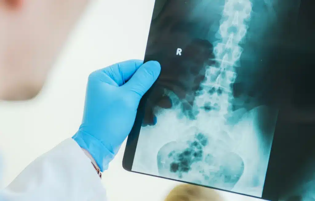
ToggleA spine fusion X-ray is a specialized imaging test used to examine the spine before, during, and after spinal fusion surgery. Unlike regular spinal X-rays, which may simply show the alignment and structure of the spine, spine fusion X-rays provide detailed images that help surgeons assess the position of hardware like screws, rods, and cages.
These X-rays are vital in ensuring that the spinal segments are properly fused together, which is crucial for the long-term stability of the spine. In my 25 years as a spine surgeon, I’ve found that these X-rays are indispensable in both planning and monitoring the success of the surgery.
There are several types of spine fusion X-rays that we use at different stages of the surgery process. Initially, we take anteroposterior (AP) and lateral X-rays to establish a baseline and to plan the surgery. These images give us a clear view of the spine from different angles, allowing us to assess the alignment and detect any pre-existing conditions that might affect the surgery.
After the surgery, we often use flexion-extension X-rays to check the stability of the fusion. These X-rays are particularly useful in detecting issues like pseudoarthrosis, where the bones haven’t fully fused together.
Another key type of X-ray is taken during the surgery itself, often referred to as intraoperative X-rays. These help ensure that the hardware is being placed correctly and that everything is aligning as it should be.
X-rays are an integral part of the entire spinal fusion process. Before the surgery, we use X-rays to assess the patient’s spinal alignment and to plan the exact placement of hardware. During the surgery, intraoperative X-rays guide us in placing screws, rods, and cages with precision.
After the surgery, we take X-rays immediately to check the hardware positioning and to establish a baseline for future comparisons. Later on, we continue to take X-rays to monitor the progress of the fusion.
In my experience, these follow-up X-rays are crucial for ensuring that the fusion is progressing as expected and that there are no complications, such as hardware loosening or movement between the fused segments.
When reviewing spine fusion X-rays, we look for several key indicators to ensure that the surgery was successful. Before the surgery, we check the alignment of the spine and look for any signs of degeneration, fractures, or other abnormalities.
During the surgery, our main focus is on the placement of the hardware—making sure that everything is aligned correctly and securely. After the surgery, we continue to monitor the hardware, looking for any signs of movement, loosening, or misalignment.
We also assess the progress of the bone fusion, which is critical for the long-term stability of the spine. If the X-rays show any signs of pseudoarthrosis or other complications, we may need to take additional steps to address these issues.

Preparing for a spine fusion X-ray is relatively straightforward, but there are a few important steps to keep in mind. First, patients should remove any metal objects, such as jewelry, that might interfere with the X-ray images.
It’s also important to inform the radiologist about any implants or previous surgeries, as this can affect how the X-ray is performed. In some cases, particularly for women of childbearing age, a pregnancy test might be necessary before the X-ray.
During the procedure, patients need to remain still to ensure clear images. The radiologist or technician will guide them through the process, which usually only takes a few minutes. After the X-ray, patients can resume their normal activities, but they should follow any specific instructions provided by their doctor.
While X-rays are a valuable tool in monitoring spinal fusion, repeated exposure to radiation is not without risks. The cumulative effect of radiation from multiple X-rays can increase the risk of developing certain types of cancer over time.
However, in my practice, we always strive to minimize this risk by using the lowest effective dose of radiation and limiting the number of X-rays to what is absolutely necessary. We also consider alternative imaging methods, such as CT scans or MRIs, especially when detailed images are needed without additional radiation exposure.
For patients concerned about radiation exposure, it’s important to discuss these risks with their doctor and explore all available options.
X-rays are a reliable way to monitor the success of a spinal fusion, but they do have their limitations. One of the primary indicators of a successful fusion is the absence of movement between the fused segments, which we can observe on flexion-extension X-rays.
Additionally, the integration of bone grafts and the stability of the hardware are key factors that we assess through X-rays. However, X-rays may not always show the full picture, particularly when it comes to soft tissues or early stages of bone fusion.
In such cases, we may recommend additional imaging, such as a CT scan, to get a more detailed view.
Understanding your spine fusion X-rays can be challenging, but with a little guidance, it becomes much easier. The first step is to familiarize yourself with the basic anatomy of the spine and the hardware used in your surgery.
When reviewing the X-rays, look for the alignment of the vertebrae and the positioning of the screws, rods, and cages. Your doctor will explain what these images mean and how they relate to your recovery. If you have any concerns about what you see, don’t hesitate to ask for clarification.
Keeping a copy of your X-ray reports and images can also be helpful for future reference.
If your spine fusion X-ray shows potential issues, such as hardware loosening or incomplete fusion, it’s important to address these concerns immediately. Contact your surgeon to discuss the findings and determine the next steps.
In some cases, additional imaging or tests may be necessary to get a clearer picture of what’s happening. Depending on the severity of the issue, treatment options could range from physical therapy to revision surgery.
The key is to act quickly to prevent further complications and to ensure the best possible outcome.
While X-rays are the most common method for monitoring spinal fusion, there are alternatives. CT scans provide more detailed images of bone structures and are particularly useful for assessing the integration of bone grafts.
MRIs, on the other hand, are excellent for evaluating soft tissues, nerve roots, and the spinal cord. These alternatives can be especially valuable when X-rays are inconclusive or when we need to reduce radiation exposure.
Discussing these options with your surgeon can help you choose the best imaging method for your situation.
GET IN TOUCH +
285 Sills Road
Building 5-6, Suite E
East Patchogue, NY 11772
(631) 475-5511
184 N. Belle Mead Road
East Setauket, NY 11733
(631) 675-6226
GET IN TOUCH +
285 Sills Road
Building 5-6, Suite E
East Patchogue, NY 11772
(631) 475-5511
184 N. Belle Mead Road
East Setauket, NY 11733
(631) 675-6226
SUBSCRIBE TO OUR NEWSLETTER +
Send us a Google review. Click this link and let us know how we did!
Review us on Yelp too.
© 2022 - 2023 Long Island Neuroscience Specialists. All Rights Reserved
Designed & Developed by Vibe Branding

Designed & Developed by Vibe Branding