Table of Contents
ToggleHave you been suffering from back pain for a long time now? Or is it just that your doctor recommended that you get an X-ray done? Either way, we know what you’re going through.
Spinal X-rays are a common procedure used to diagnose problems in the spine and nearby bones, like disc injuries or tumors.
We want to help you understand exactly what happens during a spinal X-ray so that you can make an informed decision about whether it’s right for you.
A spinal X-ray is a diagnostic test that uses radiation to create images of the spine. X-rays are a type of radiation that can pass through the body and be detected on film. They are used to diagnose injuries, infections, tumors and other abnormalities.
A spinal X-ray may be performed if you have pain in your back or neck area; have had a recent fall; have had surgery on your spine; are pregnant (especially during the first trimester); or have an infection in any part of your body.
A spinal X-ray is a diagnostic test that uses radiation to create images of the spine. X-rays are a type of radiation that can pass through the body and be detected on film.
They are used to diagnose injuries, infections, tumors and other abnormalities.
A spinal X-ray may be performed if you have pain in your back or neck area; have had a recent fall; have had surgery on your spine; are pregnant (especially during the first trimester); or have an infection in any part of your body.

If you have back pain and are not sure what it is, you should get a spinal X-ray.
Spinal X-rays are also taken when you are having a lumbar puncture (spinal tap). This can be done to diagnose many diseases that affect the spine like infections or tumors in the brain or spinal cord.
If your doctor suspects that there is something wrong with your spine but wants to know more about it before making a diagnosis, he may order an MRI scan of the spine instead of ordering X-rays because MRIs give better images than regular X-rays do.
Some people who have had strokes or other problems with their brain may need ultrasound scans as well so doctors can see how much damage has been done by these conditions on soft tissue like muscles and nerves around the brain region where they occurred.
Before your X-ray, you should make sure that you don’t eat or drink anything for at least six hours before your appointment time. You also need to ensure that you do not wear any metal objects, including jewelry and hairpins.
Moreover, ensure that all clothes that you are wearing have no metal buttons or zippers on them; if they do then remove them before going in for your X-ray scan.
If possible take off shoes with buckles made from metals as well as belts if they have buckles made from other materials than leather ones (like plastic).

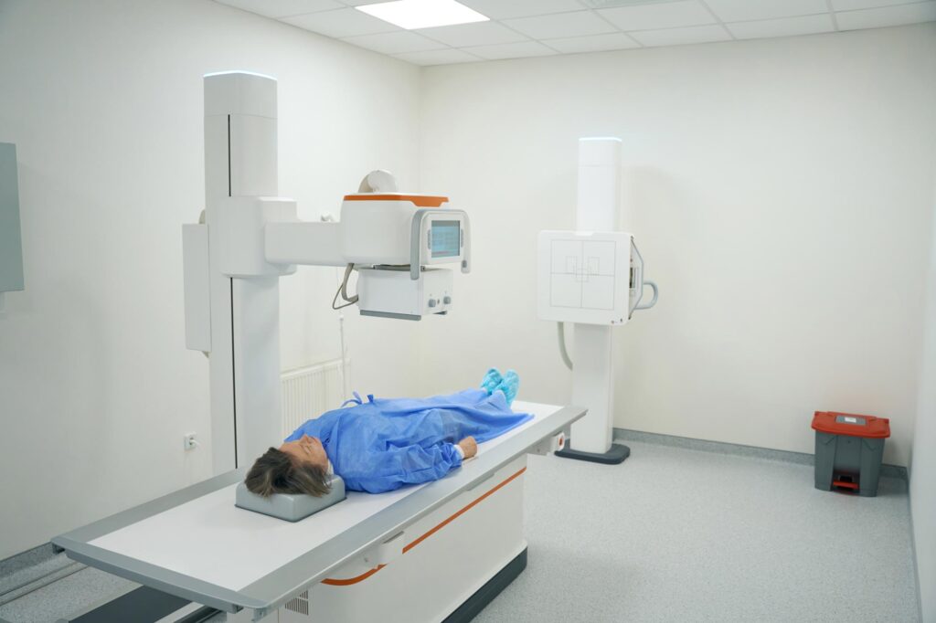
When you go for a spinal X-ray, you will be asked to lie down on a table. Your spine will be exposed and the X-ray machine is positioned behind your back.
The radiologist will take the X-ray images and they may ask you to move slightly in different positions so that they can get clear views of all parts of your spine.
Afterwards, the patient is asked to wait until all X-rays are developed (this can take several minutes).
Once this process is complete, these images are studied by doctors who then determine whether or not surgery or other treatment options are needed based on these findings.
A spinal X-ray takes about 20 to 30 minutes. You’ll need to remove any metal objects from your body, including jewelry and piercings. You should also wear loose-fitting clothes that are easy to take off.
After you arrive at the hospital or imaging center, you will be asked to undress from the waist down and put on a gown so your lower half will be visible when the X-ray is taken. You may have to wear a lead apron over your chest area if there is any concern about radiation exposure during an X-ray.
Next, you’ll lie down on an examination table with your head facing toward the machine’s viewing screen. Then you’ll be asked to hold still while the technician takes several pictures of your spine in different angles using an X-ray beam directed at specific areas of interest within the vertebral column.
Spinal X-rays are an important part of your health care, and we want you to feel safe and well-informed about the process.
In order to get the most accurate reading possible, we need to take an X-ray of your spine—but there’s no need to worry: the procedure is quick, painless, and has no lasting side effects.
If you have any concerns about this or any other aspect of your treatment at Long Island Neuroscience Specialists, please don’t hesitate to ask one of our staff members for more information.
X-rays are a form of radiation that can be used to see inside your body. They’re also called radiographs.
X-rays are used for many different reasons, including to:
In conclusion, a spinal X-Ray is a diagnostic test that is used to check the spine for fractures, tumors and other abnormalities. It’s usually performed after an injury or fall to rule out any serious problems with your vertebrae.
If your back pain is making your life unbearable, a trip to the doctor is warranted. The sooner they ascertain the cause of your pain and take the necessary steps to cure it, the better.
An X-Ray could be an important part of finally diagnosing you and helping you make progress towards treating your back problems.
GET IN TOUCH +
285 Sills Road
Building 5-6, Suite E
East Patchogue, NY 11772
(631) 475-5511
184 N. Belle Mead Road
East Setauket, NY 11733
(631) 675-6226
GET IN TOUCH +
285 Sills Road
Building 5-6, Suite E
East Patchogue, NY 11772
(631) 475-5511
184 N. Belle Mead Road
East Setauket, NY 11733
(631) 675-6226
SUBSCRIBE TO OUR NEWSLETTER +
Send us a Google review. Click this link and let us know how we did!
Review us on Yelp too.