Table of Contents
ToggleSpinal fusion surgery is a procedure where two or more vertebrae in your spine are permanently joined together. This surgery is often necessary when movement between these bones causes pain or instability, which can result from conditions like degenerative disk disease, scoliosis, or a herniated disk.
By fusing the vertebrae, we aim to eliminate this painful movement and restore stability to your spine.
In my 25 years of experience as a spine surgeon, I’ve seen how crucial it is for patients to understand the reasons behind this surgery.
Knowing why spinal fusion is recommended can ease some of the anxiety that comes with the procedure. For example, when spinal fusion is used to correct scoliosis, it’s not just about reducing pain but also about reshaping the spine to prevent further curvature.
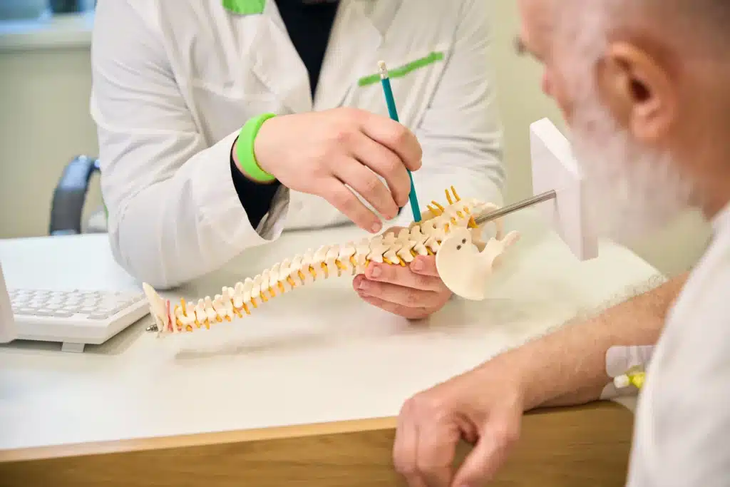
These decisions are made after careful consideration of imaging tests like X-rays and MRIs, which help us pinpoint the exact cause of the pain. Understanding these aspects is essential, and that’s where spinal fusion surgery pictures come in handy, offering a visual explanation that complements our discussions in the consultation room.
Understanding the stages of spinal fusion surgery can help demystify the procedure. The surgery begins with pre-operative preparations, which include imaging tests and marking the surgical site. These steps are crucial for ensuring accuracy during the operation.
The next stage is the actual surgical procedure, which varies depending on the approach. Whether the surgery is done from the back (posterior), the front (anterior), or the side (lateral), each method has its own set of steps, and seeing these processes through spinal fusion surgery pictures helps patients visualize what’s happening to their bodies.
The surgery itself involves making an incision, preparing the bone graft, and fusing the vertebrae. We often use metal plates, screws, or rods to hold the bones together, ensuring they heal as one solid unit. Each stage is methodical and precise, aimed at achieving the best possible outcome.
Finally, the incision is closed, and the patient is taken to the recovery room, where the immediate post-operative care begins. By breaking down the surgery into these stages and supporting it with images, we can provide a clear and comprehensive explanation that helps patients feel more prepared and confident about their surgery.
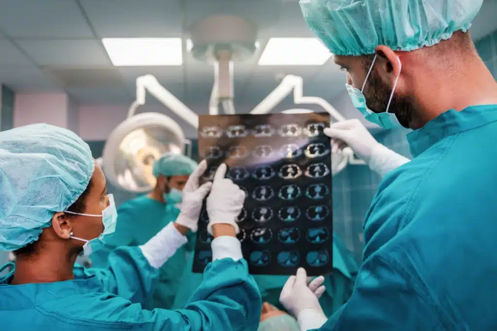
The operating room setup for spinal fusion surgery is both complex and fascinating. As you can imagine, this environment is designed for precision and safety. The room is equipped with state-of-the-art tools and technologies, all of which are essential for a successful surgery.
Spinal fusion surgery pictures of the operating room can show you what to expect, from the placement of surgical instruments to the position of the surgical team. The patient is usually placed under general anesthesia, which means you won’t feel anything during the procedure.
Depending on the approach, you might be lying on your back, stomach, or side. The surgical area is carefully prepared and sterilized to prevent infection. We use retractors to gently hold back muscles and tissues, giving us clear access to the spine.
These visual aids are not just informative; they’re also reassuring, helping you understand the level of care and precision involved in the surgery.

Spinal fusion surgery isn’t a one-size-fits-all procedure; there are different types depending on your specific condition and the part of the spine being treated. The most common types include anterior spinal fusion, posterior spinal fusion, and lateral spinal fusion.
Each approach has its own advantages and is chosen based on factors like the location of the problem and the overall health of the patient. Spinal fusion surgery pictures can effectively illustrate these different approaches, helping patients understand which method will be used in their surgery.
For instance, anterior spinal fusion involves accessing the spine from the front, often through the abdomen. This approach is commonly used for lumbar fusions and is ideal for certain types of spinal instability.
Posterior spinal fusion, on the other hand, is done from the back and is frequently used for conditions like spinal stenosis. Lateral spinal fusion is less common but is sometimes used for specific cases where the spine needs to be accessed from the side.
By providing images of each approach, we can help you visualize the procedure and understand why a particular method is recommended for your case.
One of the most valuable aspects of using spinal fusion surgery pictures is the ability to show before-and-after results. These images can be incredibly reassuring, helping you visualize the potential outcomes of your surgery.
Immediately after the procedure, the spine will look different on X-rays and other imaging scans. The fused vertebrae will appear as a single solid bone, often with visible hardware like screws or plates holding everything in place.
In the weeks and months following surgery, additional images can show the healing process. It’s common to see significant improvement in spinal alignment and stability, especially in cases where the surgery was performed to correct deformities like scoliosis.
These images also help in monitoring the progress of the fusion, ensuring that the bones are healing as expected. While every patient’s results are unique, seeing these images can provide a clearer understanding of what to expect from your own recovery.
As with any surgery, there are risks associated with spinal fusion. These can include infection, hardware complications, and in some cases, fusion failure, also known as pseudarthrosis. Spinal fusion surgery pictures can play a crucial role in identifying and understanding these risks.
For instance, an image might show the early signs of an infection, such as redness or swelling around the incision site, allowing for prompt intervention. Hardware complications, such as loosening screws or shifting plates, can also be identified through post-operative images.
Understanding these risks visually helps patients stay informed and vigilant during their recovery. While these complications are relatively rare, being aware of them—and knowing what to look for—can significantly improve your overall experience and outcome.
It’s always important to have an open line of communication with your surgeon, and having visual references can make those conversations more effective and reassuring. Want to know more about spinal fusion surgery Click Here
Spinal fusion surgery has a profound impact on the anatomy of the spine, and understanding these changes is essential for both patients and caregivers. When two or more vertebrae are fused, the mobility of that section of the spine is permanently altered.
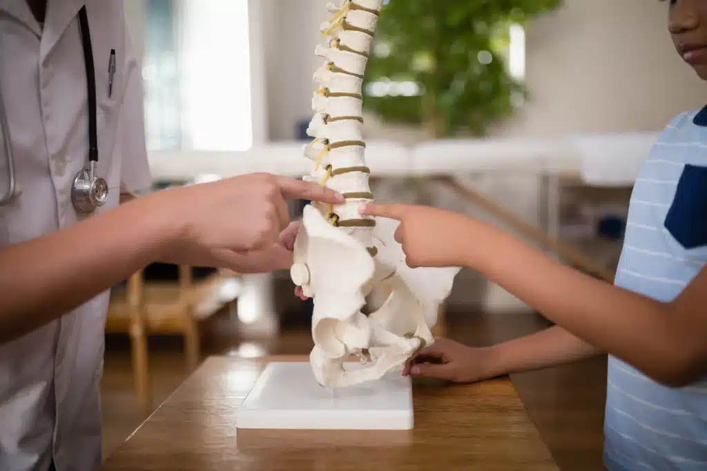
Spinal fusion surgery pictures are invaluable for showing how the anatomy changes post-surgery. Before the fusion, the vertebrae may have been misaligned or unstable, contributing to pain and other symptoms.
After the fusion, the spine is stabilized, and the previously mobile segments are now solidified into one unit. This change in anatomy also affects the surrounding vertebrae, which may experience increased stress as they compensate for the lack of movement in the fused section.
Over time, this can lead to wear and tear on these adjacent segments, which is something we monitor closely during follow-up visits. Understanding these anatomical changes through images can help you anticipate how your spine will function post-surgery and what to expect in terms of long-term outcomes.
Recovery from spinal fusion surgery is a journey, and understanding what to expect can make the process smoother and less stressful. The first few days after surgery are often the most challenging, as your body begins to heal from the procedure.
During this time, you’ll likely stay in the hospital for close monitoring. Spinal fusion surgery pictures of the early recovery phase can help you visualize the environment and the types of care you’ll receive, such as IV pain management and the use of a back brace to support your spine.
As you begin to regain strength, physical therapy becomes a crucial part of your recovery. Images of therapy sessions, exercises, and mobility aids can guide you through this phase, showing you how to move safely and effectively.
It’s important to follow your therapist’s instructions closely, as proper movement is essential for a successful fusion. Over the weeks and months that follow, your activity level will gradually increase, and you’ll be able to return to more normal activities.
These visual guides can help you track your progress and stay motivated throughout the healing process.

Visual aids are incredibly powerful tools for understanding complex medical procedures like spinal fusion surgery. Spinal fusion surgery pictures provide a clear and detailed look at each stage of the process, from pre-surgery preparations to long-term recovery.
For many patients, seeing these images can help demystify the surgery, making it easier to grasp what will happen during each step of the procedure. In my practice, I’ve found that using images to explain spinal fusion not only enhances patient understanding but also reduces anxiety.
When patients can see exactly what will happen, they feel more prepared and confident in their decision to undergo surgery. This visual approach is particularly valuable for explaining the risks and benefits of the surgery, as well as setting realistic expectations for recovery.
Whether you’re a patient preparing for surgery or a caregiver supporting a loved one, these images can provide the clarity and reassurance needed to navigate the spinal fusion journey.
If you’re looking for more information or support, there are many resources available to help you on your journey. At Long Island Neuroscience Specialists, we have a wealth of experience in spinal fusion surgery, and our team is here to guide you every step of the way.
You can find more detailed information about our services and our team of specialists on our about page, where you’ll learn about our 25 years of experience in the field. For additional support, consider joining online forums or support groups where you can connect with others who have undergone spinal fusion surgery.
These communities can be a great source of advice, encouragement, and shared experiences. We also recommend exploring educational videos and animations that further explain the spinal fusion process. These resources, combined with the visual aids provided in this article, can help you make informed decisions and feel more confident as you move forward with your treatment.
Preparation for spine surgery disc replacement involves several steps. Patients undergo comprehensive evaluations, including imaging tests and possibly nerve conduction studies. Lifestyle adjustments, such as quitting smoking and managing weight, are crucial.
Discussing all medications with your healthcare provider is essential, as some may need to be discontinued before surgery. Understanding the procedure, its risks, and benefits is vital, and having a support system in place for postoperative care can aid in a smoother recovery.

GET IN TOUCH +
285 Sills Road
Building 5-6, Suite E
East Patchogue, NY 11772
(631) 475-5511
184 N. Belle Mead Road
East Setauket, NY 11733
(631) 675-6226
GET IN TOUCH +
285 Sills Road
Building 5-6, Suite E
East Patchogue, NY 11772
(631) 475-5511
184 N. Belle Mead Road
East Setauket, NY 11733
(631) 675-6226
SUBSCRIBE TO OUR NEWSLETTER +
Send us a Google review. Click this link and let us know how we did!
Review us on Yelp too.